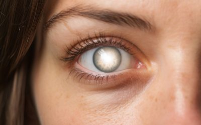
Normalmente, los ojos son estructuras que tendemos a tomar como garantizado hasta que comienzan a deteriorarse. El sentido de la vista a través de los ojos, es el que permite al hombre conocer el medio que lo rodea, relacionarse con sus semejantes, e interpretar información sobre colores, formas, distancias, posiciones y movimientos de los objetos.
Muy pocas personas conocen las partes del ojo y menos aún el nombre que tienen cada una de estas partes. Con frecuencia, encuentro en el consultorio pacientes que se confunden al contarme que tienen una catarata en vez de un pterigio, o que fueron operados de la córnea en vez de la retina; y ello es quizás porque no le damos la importancia adecuada a los ojos.
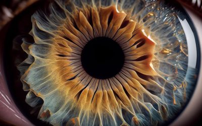
Algunos conocimientos sobre las partes del ojo permiten reconocer dónde existe un dolor y la gravedad o no del mismo. Comencemos por decir que el ojo es un órgano par y simétrico, especializado para percibir luz. Mide aproximadamente 25 milímetros y está conformado por tres capas primarias que a su vez se subdividen.

1.CAPAS DEL OJO
1.1. Capa Externa o fibrosa: es la membrana externa del ojo formada por la esclerótica, la córnea y la zona muscular donde se pegan los músculos del ojo.
– La esclerótica es la parte blanquecina externa que impide el paso de luz al ojo; está formado por fibras de colágeno y es muy resistente, lo que le da forma y protección al globo ocular.
– La córnea es la parte frontal del ojo y además el único tejido transparente del cuerpo, no tiene vasos sanguíneos, tiene 0,5 milímetros de espesor y está formada por colágeno y células llamadas “queratocitos”.
– La zona muscular sobre la esclerótica, es el área donde se pegan los seis músculos que muevan al ojo: cuatro músculos rectos que mueven el ojo hacia arriba, hacia abajo y hacia los costados; y dos músculos oblicuos que permiten el movimiento circular.
1.2. Capa Media o vascular: es la capa con mayor cantidad de vasos sanguíneos. Está formada por la coroides, el iris y la pupila.
– La coroides, es la membrana media del ojo que contiene muchos vasos sanguíneos que nutren la retina.
– La pupila o “niña” es la perforación central de la capa media, que permite la entrada de luz al ojo.
– El iris es la membrana anular anterior de la capa media, y su color varía según la cantidad y naturaleza de células pigmentarias que contenga. Está formada por fibras musculares radiales y circulares, que al moverse determinan la dilatación (midriasis) o la contracción (miosis) de la pupila.
1.3. Capa Interna o nerviosa: también llamada retina, está formada por millones de fibras nerviosas que originan el nervio óptico. Las células de la retina pueden dividirse en conos (encargados de la visión de colores) y bastones (sensibles a la intensidad de luz).
– La retina en su parte posterior presenta un circulo lleno de fibras nerviosas que es la papila o nervio óptico, y una mancha amarilla llamada mácula lútea.
– La papila o punto ciego es el lugar de la retina que es insensible a la luz porque no posee conos ni bastones.
– La mácula lútea es una región que tiene en su centro una depresión o fóvea donde se halla la mayor cantidad de células sensoriales responsables de la visión; por eso es considerada la zona de mayor agudeza visual.

2. MEDIOS TRANSPARENTES DEL OJO
Además de las capas del ojo, por dentro el sistema visual tiene estructuras transparentes muy especializadas formados por el cristalino, el humor acuoso, el humor vítreo.
2.1. Cristalino: Es un lente biconvexo, elástico, incoloro y trasparente, que se ubica inmediatamente por detrás del iris, gracias a este lente los rayos de luz provenientes del exterior se enfocan en la retina. El cristalino divide el globo ocular en dos compartimientos, uno anterior que contiene el humor acuoso, y otro posterior, que contiene el humor vítreo.
2.2. Humor acuoso: es un líquido incoloro y transparente, formado en su mayor parte por agua (98%). Se aloja en el compartimiento anterior del globo ocular.
2.3. Humor Vítreo: también llamado cuerpo vítreo, es una masa transparente y gelatinosa que llena el compartimiento posterior entre el cristalino y la retina.
3. ANEXOS DEL OJO
El ojo también depende de todas aquellas estructuras que lo rodean, encargados de protegerlo y añadirle movimiento; ellos son las cejas, los párpados, la conjuntiva, el aparato lagrimal y los músculos:
3.1. Cejas: estructuras arqueadas, cubiertas de pelo, ubicadas a 2 centímetros arriba de los ojos, son una de las partes del rostro más importantes a nivel estético y dinámico. Su función es la de proteger a los ojos de la transpiración que se desliza por la frente, y expresar sentimientos junto con los ojos a través de su movimiento.
3.2. Párpados: son dos pliegues de piel musculo mucosos, superior e inferior, que se extienden por delante del ojo. El párpado superior es más desarrollado y movible que el inferior. Ambos cumplen una función de protección contra los objetos externos y contra los excesos de iluminación.
3.3. Conjuntiva: es una delgada membrana mucosa y trasparente que cubre la parte blanca o esclerótica del ojo y la parte posterior de los párpados.
3.4. Aparato Lagrimal: consta de una glándula lagrimal, de conductos lagrimales y de un órgano reservorio de lágrimas llamado saco lagrimal.
3.5. Músculos: los músculos del ojo son seis, 4 rectos: superior, inferior, externo e interno; y 2 oblicuos: mayor y menor.

Parts of the Eye
Eyes are structures that we usually take for granted until they begin to deteriorate. The sense of sight allows humans to know their surroundings, relate to other humans and interpret information about the color, shape, distance, position and movement of objects.
Very few people are aware of the parts that make up their eyes, not to mention the name of each of those parts. In my clinic, I often find patients who tell me that they suffer from cataract, when what they actually have is pterygium, or that they underwent a corneal surgery instead of a retinal surgery; and this might be a consequence of not giving our eyes the importance they deserve.
Some knowledge about the parts of the eye may be helpful in recognizing the exact location of pain and its seriousness. Let’s start by saying that the eye is a symmetrical, even organ made to sense light. An eye has an average size of 25 mm and is made up of three primary layers, which, in turn, are subdivided into other sections.
1.LAYERS OF THE EYE
1.1. External or fibrous tunic: refers to the outer layer of the eye consisting of the sclera, the cornea and the muscular area where the tendons of the eyeball are attached.
– The sclera is the external white part of the eye, which prevents light from passing through the eye. The sclera is made up of collagen fibers and is highly resistant, offering protection and giving the eyeball its shape.
– The cornea is the front part of the eye and is the only transparent tissue in our body. It is vessel-free, has a thickness of 0.5 mm and is made up of collagen and cells called keratocytes.
– The muscular area over the sclera is where the tendons of the six extraocular muscles responsible for eyeball movement attach: four rectus muscles move the eye up, down left and right, and two oblique muscles are responsible for circular movement.
1.2. Vascular or middle tunic: the vascular tunic is the layer with the highest number of blood vessels. It consists of the choroid, the iris and the pupil.
– The choroid is the middle tissue of the eye and it contains many blood vessels to nourish the retina.
– The pupil is a central hole in the middle tunic which allows light to enter the eye.
– The iris is the anterior annular tissue in the middle layer. Its color depends on the amount and nature of pigment cells. It consists of radial and circular muscular fibers whose motion causes the dilation (mydriasis) or contraction (myosis) of the pupil.
1.3. Internal or nervous tunic: also known as the retina, this layer is composed of millions of nerve fibers which make up the optic nerve. Retinal cells are divided into cones (in charge of color vision) and rods (which respond to light intensity).
– In the back of the retina we find the optic disc, a circle filled with nerve fibers, as well as a yellow spot called macula lutea.
– The optic disc or blind spot is the only area of the retina that is not sensitive to light because there are no rods or cones.
– The macula lutea features a central depression, known as the fovea, where the highest number of sensory cells in charge of vision are located; this is why this area is where highest visual acuity is found.
2. TRANSPARENT STRUCTURES OF THE EYE
In addition to the layers of the eye, the interior of the visual system features highly specialized transparent structures: the lens, the aqueous humor and the vitreous humor.
2.1. Lens: The lens is a biconvex, elastic, colorless and transparent structure, located immediately after the iris. Thanks to the lens, light entering into the eyes is focused on the retina. The lens divides the eyeball into two segments: the anterior segment, which contains the aqueous humor, and the posterior segment, which contains the vitreous humor.
2.2. Aqueous humor: refers to a colorless, transparent fluid largely made up of water (98%). It is located at the anterior segment of the eyeball.
2.3. Vitreous humor: also known as the vitreous body, it is a transparent and gelatinous mass that fills the space between the lens and the retina in the back of the eye.
3. ACCESSORY STRUCTURES OF THE EYE
The eye also depends on all those structures that surround it, which offer protection and add movement to it. These structures are the eyebrows, the eyelids, the conjunctiva, the lacrimal apparatus and the muscles:
3.1. Eyebrows: eyebrows are arched structures covered by hair and are located 2 cm above the eyes. Dynamically and aesthetically speaking, eyebrows are one of the most important parts of the human face. Their functions are protecting the eyes against forehead transpiration and moving to complement emotional expression along with the eyes.
3.2. Eyelids: eyelids are two folds made up of skin and muscle, lined with mucous membrane and located above and under the eyeball. The upper eyelid is more developed and movable than the lower eyelid. Both eyelids protect the eyes from foreign bodies and excessive light.
3.3. Conjunctiva: the conjunctiva is a thin, clear and mucous membrane that covers the white part or the sclera of the eye and the inner side of the eyelids.
3.4. Lacrimal apparatus: consists of the lacrimal gland, the lacrimal ducts and the lacrimal sac, which serves as a reservoir for overflow of tears.
3.5. Muscles: there are six eye muscles: 4 rectus muscles –superior, inferior, medial and lateral– and 2 oblique muscles: superior and inferior.